no eosinophilia and no symptoms – no investigation or treatment required.
eosinophilia – perform stool microscopy for ova cysts and parasites (OCP) followed by directed treatment.
If no documented pre-departure albendazole therapy, depending on local resources and practices there are two acceptable options:
empiric single-dose albendazole therapy (age >6 months, weight <10kg; 200mg; ≥10kg; 400mg). If eosinophilia at baseline re-check in 8 weeks. If eosinophilia persists perform stool microscopy for OCP
OR
perform stool microscopy OCP followed by directed treatment. Recheck eosinophils and stool microscopy OCP at 8 weeks after directed treatment.
Refer if unable to find cause of eosinophilia.
Treat pathological helminths with albendazole (age > 6 months, weight <10kg; 200mg; ≥10kg; 400mg) for three days, except for Ascarislumbricoides, which only requires 400mg as a single dose (200mg in children >6 months and <10 kg). Mebendazole is an option for some parasites.136
Treat giardiasis with tinidazole 2g as a single dose, (50mg/kg in children, maximum 2g), or metronidazole 2g daily for three days (30mg/kg in children, maximum 2g).148
In people with positive stool microscopy, follow up with stool microscopy at 2-4 weeks after treatment and re-treat if necessary.
Refer refractory cases to an ID specialist.
Avoid albendazole (class D) and mebendazole (class B3) in pregnancy, both can be used during lactation.149
Overview
Background
Intestinal parasite infections are common in low resource and rural communities. The largest disease burden is caused by the soil-transmitted helminths (STH), with an estimated 2 billion people, or 30% of the world’s population, infected globally.150 Infections are common in tropical and subtropical areas, especially in Sub-Saharan Africa, the Americas, China and East Asia. The main species are Ascarislumbricoides, Trichuristrichiura and the hookworms (Necatoramericanus and Ancylostomaduodenale). Giardialamblia is a protozoan parasite that also commonly causes infection in these settings.
The prevalence of pathogenic stool parasites in people from refugee-like backgrounds reflects their socio-demographic and environmental circumstances, their countries of origin and transit, and availability of therapy.151,152
In a study of 26,956 African and South East Asian refugees between 1993 and 2007, at least one nematode was found on stool microscopy in 20.8% of 4,370 people who had not received pre-departure albendazole.117 In the 22,586 people who received pre-departure albendazole, only 4.7% had nematode infection.117,153 In 99 recent immigrants in New York, 40% had pathogenic parasites detected in stool.154 Australian prevalence data are summarised here, most studies have found the prevalence of pathogenic stool parasites is between 15–40%39,41,43,48,51,108,135,155– 159 and the most common pathogen is Giardia.
Screening
Previous ASID guidelines recommended stool microscopy if this was readily obtainable, or where symptoms were present.124 One stool sample will detect 90% of parasites.160
The voluntary Departure Health Check (DHC) has been implemented since 2005, and is now in place for most source countries19 for Australia’s offshore Humanitarian Programme intake. The uptake of the DHC is unclear; however, many offshore arrivals will have received albendazole as part of the DHC. The current guidelines consider the introduction of the DHC and recent data on the impact of albendazole on the prevalence and patterns of intestinal parasites.117,153[/accordion-item]
History and Examination
Most patients are asymptomatic.136 Symptoms due to intestinal parasites may include diarrhoea, cramping and abdominal pain.
Investigation
Offer all an FBE to look for eosinophilia.
People with documentation of pre-departure albendazole treatment do not require screening for faecal parasites unless they are symptomatic or if they have eosinophilia.
In people with no documentation of pre-departure albendazole there are two acceptable options, depending on local resources and practices:
Give empiric single-dose albendazole therapy (>6 months, <10kg; 200mg; ≥10kg; 400mg). This will be effective against many of the common parasites (see below). If they have eosinophilia at base line, re-check at 8 weeks post treatment. If eosinophilia unresolved, refer to specialist.
OR
Perform stool microscopy for OCP. Obtain at least one fresh or fixed specimen delivered promptly to the laboratory. If there is a delay with delivery, stool should be in preservative (SAF). Laboratories are funded through Medicare for one OCP exam in seven days.
These may be found in stool but no further action needs to be taken: Entamoeba coli
Entamoeba hartmanii
Entamoeba gingivalis
Endolimax nana
Iodamoeba butschlii
Dientamoeba fragilis Blastocystis hominis (note: rarely implicated as a pathogen, discuss with ID) Chilomastix mesnili
Trichomonas hominis [/accordion-item]
Management and Referral
Note: Albendazole is available at some refugee health and specialist hospital clinics, or on the PBS via streamlined authority for treatment of tapeworm and hookworm.
Albendazole 400mg daily for three days orally if weight ≥10kg (200mg daily if >6 months and <10kg).
Albendazole has been shown to be superior to mebendazole, but single dose treatment has suboptimal efficacy for hookworm infection.161–164 Treat any concurrent iron deficiency (see Anaemia, Iron Deficiency and Other Blood Conditions).
Round worm (Ascaris lumbricoides)
Albendazole 400mg orally if weight ≥10kg (200mg >6months weight <10kg) as a single dose.161,162 Corticosteroids are occasionally required in pulmonary ascariasis.
Whipworm (Trichuris trichiura)
Albendazole 400mg orally for three days orally if weight ≥10kg (200mg daily if >6 months and <10kg).
A three-day treatment regime had an efficacy of 83% in an RCT involving 175 children in Gabon.163 Single dose therapy has low efficacy for trichuriasis.161,164
Giardia lamblia
Treatment is with tinidazole 2g orally as a single dose (50mg/kg in children, maximum 2g), or metronidazole 2g daily for three days (30mg/kg in children, maximum 2g) (efficacy >90%).136,148
Albendazole 400mg daily for five days is probably as efficacious as metronidazole 500mg three times daily for five days, with fewer side-effects.165
Caution: Albendazole
Albendazole should be used with caution in patients who have symptoms and/or a travel history compatible with neurocysticercosis (such as epilepsy, central nervous system (CNS) symptoms, subcutaneous nodules, Taenia solium positive in faeces or serology) as treatment with albendazole alone can exacerbate CNS disease.
Follow-up
Repeat stool microscopy for OCP 2–4 weeks post therapy. Retreat if ova still present. Refer refractory cases for specialist management.
Considerations for Children, and for Pregnant and Breastfeeding Women
Albendazole is a class D drug. It should not be used in the 1st trimester of pregnancy. WHO recommends use in 2nd and 3rd trimester.166 In women who are breastfeeding pyrantel is an alternative to albendazole.
Avoid albendazole in children ≤6 months, and give 200mg dose if >6months and <10kg. Avoid mebendazole in pregnancy (class B3).
Australian therapeutic guidelines state both albendazole and mebendazole can be used during lactation.76
Seek specialist advice if uncertain, and refer children <2 years for specialist review if concerned.
Joshua S Davis, Christine Phillips, Vanessa Clifford
Recommendations
Offer blood testing for schistosomiasis serology if people have lived in/travelled through endemic countries (including Africa, parts of South East Asia and the Middle East, see text).
If serology is negative, no follow up is required.
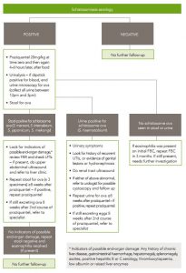
If serology is positive or equivocal:
Treat with praziquantel in two doses of 20/mg/kg, 4 hours apart, orally. (40mg/kg total, no upper limit) (EBR – A)
Perform stool microscopy for ova.
Perform urine dipstick for haematuria, and end-urine microscopy for ova if haematuria.
If positive for ova on urine or stool, evaluate further for end-organ disease with ultrasound and LFTs. See flow-chart for further details.
Seek advice from a paediatric specialist on treatment of children <5 years.
Overview
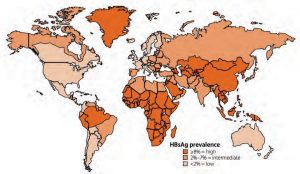
Schistosomiasis is a chronic parasitic infection caused by flukes of the genus Schistosoma. The infection is acquired through contact with infective cercariae in fresh water (e.g. swimming or bathing) that have been released by an intermediate snail host in fresh water. Schistosomiasis affects 200 million people worldwide and is estimated to cause over 200,000 deaths per year in Africa.113 Urinary schistosomiasis is caused by Schistosoma haematobium, which is found in Africa, the Middle East, Corsica, France, and intestinal schistosomiasis is caused by S. mansoni, S. intercalatum, S. japonicum, and S. mekongi, which are found in Africa, South America and South East Asia.114
Epidemiology in common countries of origin
Africa has the highest prevalence of schistosomiasis of any region, accounting for approximately 95% of affected people worldwide.113 Schistosomiasis is much less common in South East Asia, although it is prevalent in China (along the Yangtze River), the Philippines, parts of the Mekong River valley (Laos, Myanmar [Burma], Vietnam) and Indonesia. In the Middle East, there are residual foci of schistosomiasis in the Arabian Peninsula, including in Syria.
Table 6.1 shows a list of countries where schistosomiasis is endemic.
*These countries have had significant decreases in disease incidence due to WHO control programmes, but disease is still prevalent.
Seroprevalence data from post-arrival refugee screening
Many centres in Australia currently screen people from refugee-like backgrounds for schistosomiasis using serology and, in some cases, stool and urine testing. Most screening data come from African refugees, in whom schistosomiasis is common. Positive schistosomiasis serology has been found in 37% of African refugees arriving in Newcastle NSW,115 38% arriving in Hobart,116 and 12% in Melbourne.108 Fewer data are available on seropositivity in non-African refugees, with seropositivity in 9% (57/632) of Karen adults and 4.6% (23/504) in Karen children arriving in Melbourne,43 and 18% of 110 Asian refugees arriving in Darwin.39 These patients were part of population screening programmes for all new arrivals and thus are likely to be representative of the population as a whole.
Stool prevalence data from post-arrival refugee screening
In contrast to serological screening, screening based on stool microscopy is less sensitive and leads to a much lower rate of diagnosis. Western Australian data for all arrivals in 2003 and 2004, showed 3-7% of 1,633 African refugees had schistosome ova in stool, but none of 146 South East Asian refugees.41 Similarly, in a large study of 4,370 refugees arriving in Minnesota, USA, schistosomiasis was diagnosed on faecal specimens in 5.6% of African refugees but none from South East Asia.117
Rationale for screening
Schistosomiasis is a chronic disease that is generally asymptomatic until the late stages, when there is significant end-organ damage. Treatment is safe, simple and generally highly effective. The consequences of not diagnosing and treating chronic schistosomiasis range from none (in a light infection) to early mortality from portal hypertension, renal failure or bladder malignancy. There can also be substantial morbidity and long-term cost to health services as a result of intestinal polyposis, oesophageal varices, hydronephrosis, and urogenital fibrosis. Adult worms can live within venous plexuses for 20-30 years (mean 7 years) after the patient has left an endemic area. Thus, schistosomiasis meets all the usual criteria for health screening: available and effective diagnostic tests and treatment, a long asymptomatic phase, cure with early detection and treatment, and high prevalence.
History and Examination
Most infected individuals are asymptomatic and are therefore unlikely to seek treatment. Symptoms of chronic infection may include abdominal pain, bloody diarrhoea or cystitis/ haematuria, or rarely, genital sores. See ‘Management’ for evaluation of end-organ damage related to schistosomiasis.
Investigations
Examination of stool and/or urine for ova is the ‘gold standard’ diagnostic method for schistosomiasis; however, ova will only be identified if there are multiple specimens submitted and there is a high burden of egg excretion.118 It is also logistically difficult and not cost-effective to collect and process multiple stool samples on an initial health assessment visit. For these reasons, serology is the recommended screening test, with stool and/or urine microscopy for schistosoma ova only in people with positive serology.
The reported sensitivity and specificity of serology varies with the method used.119 In general, modern methods are highly sensitive and specific. For example, the Institute for Clinical Pathology and Medical Research (ICPMR), the parasitology reference laboratory for New South Wales, uses an in-house ELISA with extracted egg antigens from S. mansoni. This technique has had excellent reported results in other centres, with sensitivity for chronic infection with S. mansoni of 93.3% and specificity of 98.2%.120 However, the sensitivity is not as good for non-mansoni species, with an estimated sensitivity of 90% for S. haematobium and 50% for S. japonicum and mekongi.121
Serology can remain positive after effective treatment, or after the infection has run its course. Thus it is a highly sensitive test (for African forms at least), but cannot reliably differentiate between current and past infection.114 Most people living in endemic areas are repeatedly infected due to bathing or washing in contaminated water sources until they leave the area or effective local control measures are put in place. This, combined with the fact that worms can survive for decades, means it is likely that the majority of people from schistosoma endemic areas with positive serology have current infection.
Schistosomiasis is a differential diagnosis for unexplained eosinophilia, and screening should be completed in people of refugee-like background with eosinophilia who have spent time in endemic areas.
Management
Praziquantel (40 mg/kg in two divided doses of 20mg/kg, taken four hours apart, pregnancy category B1) has excellent cure rates in schistosomiasis acquired in Africa, South East Asia and South America and is generally safe and well tolerated.122
Doctors and nurse practitioners can prescribe praziquantel for schistosomiasis through the PBS under streamlined authority arrangements. Each 600mg tablet can be readily divided into four, allowing dosages to be delivered to the nearest 150mg, or quarter of a tablet. Use Easidose to explain medication dosing.
Table6.2: Conversion of praziquantel tablet quantities corresponding to one 20mg/kg bodyweight dose123
Body weight (kg)
Tablet Quantity
20—25
¾
26—33
1
34—41
1¼
42—48
1½
49—56
1¾
57—63
2
64—70
2¼
71—78
2½
79—86
2¾
The previous version of this124 and other guidelines recommended a higher dose of praziquantel (60mg/kg in 2-3 divided doses) for schistosomiasis acquired in Asia. However, this recommendation was based on very weak evidence, and a recent international randomised controlled trial (RCT) found that 40mg/kg resulted in comparable cure rates to 60mg/kg in patients with schistosomiasis from Africa, the Philippines and Brazil.125 Hence we recommend a uniform dose of 40mg/kg in two divided doses in all patients regardless of their area of origin.
Mild adverse effects occur with praziquantel in 5-30% of patients – these include nausea, dizziness, headache, diarrhoea and pruritus. Discuss potential side effects with patients.
These symptoms may be immune reactions to the dying worms rather than direct adverse effects of the drug.126 There is a theoretical concern of precipitating seizures in patients with neurocysticercosis (encysted Taenia solium parasites in the central nervous system). However this complication is very rare and it has not been reported in Australian refugee clinics to date. Routine cerebral imaging to exclude concurrent neurocysticercosis prior to praziquantel is not indicated. However, in a person with unexplained seizures, fundoscopy (for retinal lesions of neurocysticercosis) and cerebral imaging should be performed. Patients with possible neurocysticercosis should be referred to an ID specialist prior to praziquantel treatment.
Follow up
If serology is positive, examine stool for schistosome ova. See flow chart (figure 6.1) for treatment and follow-up recommendations. This is not as a screening test, but as a risk-stratification tool, to detect patients with high worm burdens (in whom treatment failure is more likely with a single episode of treatment). Similarly, only people with positive serology and macro- or microscopic haematuria (using dipstick urinalysis) should have their urine tested for ova.
Patients with positive stool or urine microscopy for schistosome ova should have further follow up (see figure 6.1). This includes repeat stool or end urine microscopy to document cure, and evaluation for end-organ damage, including LFTs in those with positive stool microscopy. For S. mansoni infection, people with features of hepatic impairment should have imaging with liver ultrasound to exclude ‘pipe-stem’ fibrosis, a well-recognised complication of chronic S. mansoni infection. Features of liver impairment include: any history of chronic liver disease, ascites, gastrointestinal haemorrhage, chronic hepatitis B or C infection; examination findings of hepatomegaly, splenomegaly, ascites or peripheral oedema; or positive hepatitis B or C serology, thrombocytopaenia, low albumin or raised liver enzymes. If a patient has features of liver impairment, they should be referred to a specialist ID physician.
For S. haematobium infection, all patients with ova in urine should have a physical examination for genital disease and a renal tract ultrasound to exclude bladder and renal pathology. People with macroscopic haematuria, recurrent UTIs, abnormalities on imaging or genital disease should be referred to a urologist for consideration of cystoscopy and further follow up.
After successful treatment of schistosoma infection, ova may be excreted for weeks. Therefore, delay repeat stool or urine examination to document cure for at least 8 weeks after treatment. If stool or urine remains positive for ova despite a course of praziquantel, treatment should be repeated. If stool or urine remains positive for ova more than 8 weeks after a second course of praziquantel, refer to an ID physician.
Do not repeat serology post treatment as this may stay positive for years.
Do confirm adherence to treatment as tablets and dosage are quite confusing and treatment may need to be re-explained (and re-prescribed if not taken the first time).
Considerations for Children
Children should be evaluated and treated as per the flow chart in the same way as adults (figure 6.1). Since the consequences of residual infection (e.g. growth delay, cognitive impairment) may be more significant in children, there should be a low threshold for referral to a paediatric ID specialist, especially if eosinophilia has not resolved and/or ova are still detectable in urine or stool after the first course of treatment. In addition, consultation with a specialist is recommended for children <4 years, as the safety and efficacy of praziquantel in this age group is not as well established,127 although praziquantel has been used in children as young as 12 months in mass eradication campaigns.
Pregnancy and Breastfeeding
Praziquantel is a category B1 drug for use in pregnancy. There is controversy about whether women with asymptomatic schistosomiasis should be treated during pregnancy; however, there is a risk of loss to follow up if treatment is deferred until after pregnancy. There is emerging evidence about the safety of praziquantel in pregnancy 128 and no convincing evidence of teratogenicity. The WHO now recommends that pregnant women in endemic areas are included in mass treatment programmes. We recommend that praziquantel treatment for asymptomatic schistosomiasis is withheld during the first trimester, but either offered subsequently (after discussing the risks and benefits with the patient), or administered after delivery. There is no evidence of praziquantel causing harm to infants of breastfeeding mothers, and thus we also recommend that praziquantel treatment is offered to lactating mothers where indicated.
Offer screening to all people from refugee-like backgrounds for anaemia and for other blood conditions with a full blood examination (FBE).
Offer screening for iron deficiency with serum ferritin to all children and to women of childbearing age and consider this in patients with unexplained fatigue.
Replace iron if ferritin <15µg/L and/or when clinical and haematological features indicate iron deficiency anaemia.
Educate about iron-rich diet and avoid excessive dairy intake in children
Investigate and treat causes of anaemia
Consider screening for vitamin B12 deficiency if arrival <6 months with a history of at least several years of significantly restricted food (especially meat) access e.g. patients from Bhutan, Afghanistan, Iran or the Horn of Africa; or if vegan diet.
Treat B12 deficiency if serum active B12 <35pmol/L or <reference range for children with oral or IM supplementation. Exclude concomitant folate deficiency. Consider Helicobacter pylori infection.
Overview
There are food security risks in many refugee-source countries,252 as prolonged food deprivation and inadequate access to nutritious food and clean water are common. After arrival in Australia there still may be issues of food insecurity, 253,254 poor access to healthy foods and consequent under-nutrition. Specific issues include: low weight and/or height-for-age in children, vitamin deficiencies, iron deficiency and anaemia. As well as the potential for under-nutrition, there are increasing problems with dyslipidaemia and obesity and the associated risks of developing non-communicable diseases (NCDs chapter 14). The period of early settlement is a window for health promotion about nutrition.
Anaemia (defined as a low blood haemoglobin concentration with ‘normal’ ranges depending on gender, age and pregnancy status) has been reported in adults from refugee-like backgrounds in Australia. The prevalence is estimated at 7–20%, 39,43,45,255 but is greater in young children (23–39%)43,255 (see prevalence tables for more information). Anaemia is a major cause of, or a contributor to, morbidity and mortality worldwide. In some settings up to 50% of patients with anaemia are iron deficient.256 Iron deficiency in children from refugee backgrounds in Australia and New Zealand ranges from 17% (36/216)45 to 33% (113/343).51
Iron deficiency anaemia occurs after iron stores become severely depleted. As well as the common causes in the Australian population such as pregnancy, breast feeding, menstrual blood loss, inadequate iron intake, and gastrointestinal blood loss,257 chronic infections such as hookworm, strongyloidiasis, Helicobacter pylori infection and chronic malaria can contribute. Iron deficiency, even without associated anaemia, may cause fatigue and hence affect productivity. Women of reproductive age and children are at particular risk of iron deficiency and iron deficiency anaemia because of greater iron requirements. It is important that women have adequate iron stores during their reproductive years, as iron deficiency in pregnancy increases maternal and perinatal mortality, and can cause cognitive and motor delays in children. Side effects of iron supplementation include gastrointestinal symptoms such as nausea, constipation or abdominal pain as well as the potential for overdose in children.258–262 In view of the high prevalence of anaemia and iron deficiency in people from refugee-like backgrounds, the fact that iron deficiency is easily treated, as well as published evidence for possible improvements in symptomatic fatigue in women 263 and for psychomotor development in children,264 we recommend screening for anaemia with full blood examination (FBE) in all patients and to consider excluding iron deficiency by determining serum ferritin concentrations in women of child-bearing age, children, and those with fatigue. FBE, rather than purely haemoglobin (Hb), is recommended because red cell characteristics may be useful in determining the type of anaemia, and differential white cell concentrations (e.g. eosinophils) are included.
Other haematological conditions
Congenital neutropenia. Neutrophil concentrations below normal Australian reference ranges occur in 25–50% of persons of African descent and in some groups from the Middle East.265 This is not a pathological condition but rather reflects a different ‘normal’ range. If the person seems well and has no associated clinical features like fever, gingivitis or skin infections, no further investigation is indicated.
Eosinophilia is defined as an absolute eosinophil count exceeding 0.6 x 10⁹ eosinophils/mm3 and is apparent on FBE. Eosinophilia in this population may indicate the presence of a parasitic infection (see schistosomiasis, strongyloidiasis, and other intestinal parasites). Note: single-cell intestinal parasites (e.g. giardia) do not cause eosinophilia. Other causes of eosinophilia include allergies and medications.
Inherited anaemias. These include thalassaemias, G6PD deficiency and haemoglobinopathies. These are more common in people from Africa, Asia and the Middle East. Carriers are usually asymptomatic but often have mild microcytic hypochromic anaemia.
Lead toxicity. Elevated serum lead concentrations have been reported in up to 7–25% of people of African refugee-like background266,267 and in children from the Thai-Burma refugee camps267–270 especially in those aged < 6 years, although rarely to a level requiring chelation therapy. Screening for lead toxicity in Australian refugee populations is not recommended routinely but should be considered if there are symptoms of lead toxicity such as learning, memory, behavioural or cognitive dysfunction in children271 and when anaemia is unexplained.
Coeliac disease. Although there are no Australian data available in people from refugee-like backgrounds, the rates of coeliac disease in North Africa and the Middle East are apparently similar to rates in Western countries. The disease is rare in Sub-Saharan Africa and in East Asia.272
Vitamin B12 deficiency has been reported in refugees from Bhutan, Iran, Afghanistan, Iraq and the Horn of Africa in Australia 156,273,274 especially when there is poor access to food. This does not necessarily correlate with symptoms or even with macrocytosis, and the significance of such apparent deficiency, the requirements for replacing borderline deficiency, the effects of post-migration diets and possible impacts on long-term health in those from refugee-like backgrounds are currently unclear.275
Folate deficiency is uncommon in those from refugee-like backgrounds in Australia 43,273 but should be considered in new arrivals with food insecurity. As folate deficiency is uncommon in Australia due to the availability of fresh foods and vegetables, and the fortification of some foods, testing is not recommended.
History and Examination
Symptoms of anaemia may be subtle. They include lethargy, irritability, shortness of breath, poor growth, weakness and signs of cardiac failure. There may be a past history of malaria, worm infestations, Helicobacter pylori infection, haematemesis or malaena and patients may have occult chronic diseases, such as renal failure. The family history may indicate a predisposition to haemoglobinopathy.
Consider folate or B12 deficiency in those with a history of food insecurity, vegan diets or with symptoms suggestive of cognitive deficit, neurological or neuropsychiatric problems, developmental delay, and, if FBE indicates macrocytic anaemia.
Investigations
Full blood examination (FBE) in all. This is to screen for anaemia, eosinophilia and thrombocytopenia.
Serum ferritin for children and for women of childbearing age, people with vegetarian, especially vegan diets, those with a history of food insecurity, if anaemia is unexplained, and when iron deficiency is suspected clinically or from FBE results. Note: in patients with acute and/or chronic inflammatory and some other conditions, serum ferritin levels of up to 60-100µg/L257 do not exclude iron deficiency.
Consider B12 screening in those with suggestive symptoms, macrocytosis, or deemed at risk (e.g. vegan diet). Screening is generally not required for asymptomatic individuals with an adequate diet, or for those living in Australia for >6 months and who consume meat. Request serum B12, if this is abnormal or low a serum active B12 (holotranscobalamin) should be ordered.
Management
If FBE shows microcytic anaemia and/or low ferritin consider and treat underlying causes. Consider common parasitic infections and Helicobacter pylori infection. Educate patients and families about iron-rich diets.
Replace iron as per published guidelines.257,276 There are a number of formulations of iron and the dosage depends on weight in children. Intermittent oral iron (e.g. once or twice-weekly) is also an option and may reduce side-effects. Discuss side-effects (e.g. dark stools, constipation, gastrointestinal upset), safety and storage with patients and families. Use Easidosefor picture-based dosing, if needed.
If iron-deficiency anaemia does not resolve within three months of oral iron supplementation and in those without a clear explanation for their anaemia, consider gastroscopy and colonoscopy especially in men and post-menopausal women, to exclude occult gastrointestinal bleeding associated with peptic ulceration, polyps, cancers etc.
B12 deficiency. In those with low or equivocal serum B12 concentrations, confirm deficiency by measuring active B12 (serum holotranscobalamin), and treat if the concentration is <35 (pmol/L).277 When B12 supplementation is indicated, consider and treat concomitant folate deficiency. Consider testing for helicobacter pylori infection.278,279 Replacement can be oral (cyanocobalamin 50–200 mcg daily, given between meals) or intramuscular (cyanocobalamin or hydroxycobalamin, IM 1000mcg given once.276 Dietary B12 intake following settlement in Australia is usually adequate and so it seems unlikely that ongoing supplementation is required, although this has not yet been studied.
The principles of healthy eating are universal and should be discussed with patients and their families.
Be aware of concurrent micronutrient deficiencies. These are outside the scope of this document but are summarised in the RCH guidelines.
Refer to a paediatrician if there malnutrition is suspected. Consider hospital admission if there is anaemia is severe (e.g. Hb < 6g/dl). Severely malnourished children should have anthropometry performed by a dietitian. Once the initial screen has been completed and treatment initiated, growth-monitoring should be offered. http://www.rch.org.au/uploadedFiles/Main/Content/immigranthealth/TE%20V2.htm
Asylum seekers are people who arrive in Australia and subsequently apply for protection as refugees
Depending on the person’s mode and date of arrival in Australia, living arrangements and service eligibility will vary
Visas and entitlements, including eligibility for Medicare, can change during the protection visa application process
All people found to be refugees in Australia undergo a Visa Health Check which is performed by a provider contracted by the Commonwealth Department of Immigration and Border Protection
Do not provide legal advice unless you are qualified to do so – if a person asks questions about their asylum claim, health professionals should refer them to a legal clinic or to advice and resources that have been prepared by legal services
Overview
Protection visa applications that are made in Australia are assessed by the Department of Immigration and Border Protection (DIBP) to determine whether the person legally engages the Australian Government’s protection obligations. Over recent years there have been multiple changes to how this processing occurs, and depending on the mode and date of arrival, different groups of asylum seekers have been and are processed under different systems, with different entitlements to have DIBP decisions reviewed.
Asylum seekers arriving with a valid visa (usually by plane)
Those arriving with valid entry documentation (e.g. a student visa or visitor visa) are permitted to reside in the community while their application is considered and are often provided with a Bridging Visa for this purpose (e.g. Bridging Visa A, Bridging Visa E). This group of asylum seekers are Medicare eligible even though if they may not have work rights.
Asylum seekers arriving without a valid visa (usually by boat)
Asylum seekers who come by boat on or after 1 January 2014 are transferred offshore to either Nauru or Manus Island, Papua New Guinea, to have their protection claims assessed by those countries.
Immigration detention (including alternate places of detention, immigrant transit accommodation, and immigration detention facilities)
Asylum seekers who arrived without a valid entry visa are subject to periods of immigration detention. Those who arrived before 31 December 2013 were usually detained on Christmas Island in the first instance, and then moved to mainland immigration detention facilities. While in detention facilities, healthcare is facilitated by the DIBP contracted service International Health and Medical Services (IHMS). Contracted hospitals that have reimbursement arrangements with the DIBP also provide care to people in immigration detention. People in detention are accompanied by guards to all appointments outside of the detention facility. After release from a detention facility, asylum seekers are allocated a Status Resolution Support Service Caseworker (SRSS provider). They are given a detention health discharge summary prepared by IHMS. If this is misplaced, health professionals may request a copy from the SRSS provider.
Community placement (previously known as community detention)
Some asylum seekers are released from immigration detention facilities into the community under residence determination arrangements. Placement in the community allows people to move about without being accompanied. DIBP have contracted service providers under the SRSS program to provide housing, case management support and, where appropriate, counselling for pre arrival experiences of torture and trauma. Community placement clients are not eligible for Medicare, instead IHMS is contracted by the DIBP to facilitate and pay for a specified range of health services for this group.
Living in the community post-detention
Asylum seekers may be released from detention facilities on a Bridging Visa E (BVE) to live in the community. This group are reliant on the private rental market, and receive housing and case work support from SRSS providers after they exit detention. Holders of BVEs waiting for the outcome of their protection visa application are eligible for Medicare and may have associated work rights. Some asylum seekers who have appealed a negative decision and whose case is at judicial review may be living in the community without Medicare and work rights. Medicare validity and expiry is also linked with the BVE. In circumstances where a BVE has expired due to DIBP administrative processing delays, a client remains in the community without a valid Medicare card. In these instances payment for medical services may be arranged in advance with SRSS providers through a letter of supply.
Code of behaviour
People who arrived by boat and are 18 years of age or older must sign the DIBP Code of Behaviour before they are considered for the grant of a Bridging E visa. The Code of Behaviour makes certain kinds of behaviour (over and above Australian criminal laws) potentially punishable by cancellation of that Bridging Visa, and therefore detention. The Code includes a requirement to ‘comply with any health undertaking provided by the Department of Immigration and Border Protection or direction issued by the Chief Medical Officer (Immigration) to undertake treatment for a health condition for public health purposes’.
Protection visas (permanent and temporary)
People who arrive in Australia with a valid visa then apply and are found to be owed protection, are entitled to a Permanent Protection Visa, subclass 866. This entitles holders to permanent residency and a pathway to citizenship and the ability to apply to sponsor their family.
People who arrive in Australia without a valid visa, then apply and are found to be owed protection, are entitled to a Temporary Protection Visa (TPV, subclass 745) for up to 3 years or a Safe Haven Enterprise Visa (SHEV, subclass 790) for up to 5 years. TPV and SHEV holders are eligible for Medicare for the duration of their visa. On either form of temporary visa, it is not possible to become a citizen, or to sponsor overseas family members to come to Australia. The Safe Haven Enterprise Visa allows you to then apply for a limited range of other kinds of visas if you work or study for 3.5 years in designated regional areas.
Important considerations for the health care of people seeking asylum
Some asylum seekers living in the community are ineligible for Medicare. Asylum seekers may be eligible for assistance with health care and income support through the Status Resolution Support Services (SRSS) program. Those who are Medicare ineligible and not eligible for SRSS rely on specialist asylum seeker health services and other, often pro bono, services.
Containing the cost of care will be important as asylum seekers may face restrictions on their rights to employment, income support and other benefits.
Asylum seekers may not have undergone the Immigration Medical Examination offshore, but will do so in Australia as part of their application for permanent protection.Practice tips and considerations for working with people seeking asylum
Mental distress and suicide risk
“Asylum seekers can spend years in the community awaiting final determination of their case. The fear of being returned home, coupled with isolation and destitution, can be overwhelming. Statements of suicidal thought by an asylum seeker should always trigger an action response plan, which may include provision of enhanced support or calling in the specialist mental health crisis team.”1
Impact of poverty
“Many asylum seekers may eat poorly and frugally. Over winter, almost all asylum seekers economise on heating, and in summer on cooling. GPs should be aware of food banks in their community and the local charities that provide clothing and other essential items.”1
Affordability of medications
“Asylum seekers with chronic diseases are often faced with choosing between medications. GPs should assist them in decision-making about which medications to prioritise and, where possible, should prescribe the cheapest medication in its class or for the therapeutic purpose.”1
Confidentiality and interpreters
Be aware that a telephone interpreter may be preferred by the patient for confidentiality reasons – especially if they are from a small community or language group
The patient’s name does not need to be given to interpreting services. You can state that this is confidential and this can speed up the process. In circumstances where the client is extremely concerned about confidentiality, offer to call the client by another name during the consultation and book an interstate interpreter if possible.
Status Resolution Support Services (SRSS) Programme
Pro-bono medical services
References
Phillips, 2014, ‘Beyond Resettlement: Long-term care for people who have had refugee-like experiences’, Australian Family Physician, Volume 43 Issue 11
Assess diabetes and CVD risk earlier for those from regions with a higher prevalence of non-communicable diseases (NCDs) or an increased BMI or waist circumference.
Although this chapter does not specifically refer to children we recommend recording body mass index (BMI) and blood pressure (BP) in all and offering management if abnormal.
Overview
Non-communicable diseases (NCDs) include CVD, diabetes, respiratory diseases such as chronic obstructive pulmonary disease (COPD), musculoskeletal diseases and some cancers. There are few published data on the prevalence of NCDs in people from a refugee-like backgrounds in Australia. Some studies report a higher rate of NCDs in some refugee populations.280 There is published evidence that people from refugee-like backgrounds may have an increased prevalence of diabetes,280–282 hypertension,282–285 dyslipidaemia,156,282 musculoskeletal disease, chronic respiratory disease,280 obesity or overweight274,280,283,284,286 and CVD,282,287 compared to the population of country of settlement.
Conversely, some studies show a reduced prevalence of NCDs in some groups of immigrants living in high-income countries (HICs) compared to the long-term residents: the ‘healthy migrant effect.’288,289
The rates of death related to NCDs are increasing in Australia290,291 and internationally particularly in low- and middle-income countries (LMIC), including refugee-source countries.292,293 Premature death attributable to NCDs in LMIC occurs at a far higher rate than in Australia.294
The major potentially treatable risk factors for the development of NCDs are poor nutrition and low physical activity, which pre-dispose to obesity, excessive alcohol intake, cigarette smoking and hypertension.
There may be additional explanations for the increased prevalence of NCDs. For immigrants moving from a low- or middle-income country to a high-income country (including people from a refugee-like background) these include abdominal obesity, dyslipidaemia, genetic and environmental factors.295–297 Experiences of prolonged poverty, forced migration, time spent in refugee camps or similar circumstances may also be potential risk factors for developing NCDs. For example, perinatal deprivation or malnutrition is considered to pre-dispose to diabetes and to other NCDs. 298,299 Post-traumatic stress disorder (PTSD) and depression are associated with hypertension and hyperlipidaemia.300 People from refugee-like backgrounds with NCDs may also be at risk of complex and multiple comorbidities due to poor socioeconomic conditions and concomitant mental illness, as well as inadequate access to preventive and chronic disease care.
Nutrition and obesity
There are few studies investigating risk factors for obesity in people from refugee-like backgrounds specifically but low socioeconomic status, food insecurity, high intake of sugar-sweetened drinks, body image and inappropriate beliefs related to food, stressful family life and depression are all risk factors for obesity in the general community, including those from refugee-like backgrounds.288,301,302 Some refugee-background children reportedly assimilate quickly to less healthy Australian diets,303 and cultural groups may value childhood obesity as a sign of health and success.304 Other refugee-background communities feeling overwhelmed by food choices305 and have limited knowledge and access to information about healthy food options in Australia.301
Physical activity
There are few published data on physical activity in people from refugee-like backgrounds. Amongst culturally and linguistically diverse communities, some refugees were less likely to be active or to consider physical activity due to PTSD.306
Diet and exercise requires individualised and culturally sensitive approaches.306
Tobacco, alcohol and other substances
Little is known about the use of substance in refugee-background communities in Australia.307,308 A recent review of substance use in people from refugee backgrounds in the USA suggested a ‘refugee paradox’ much like the ‘healthy migrant effect.’309 Nevertheless drug, tobacco and alcohol issues have been identified as specific health issues of concern following consultations with refugee groups in Australia.15,310 There are no data on the prevalence of smoking in people from a refugee-like background, and reported prevalence in immigrants varies according to gender, time since migration and country of origin.311 There are reports of increased smoking behaviour and heavy alcohol use in people from a refugee-like background who experience PTSD.308 Tobacco intake via a water pipe has been reported in those from the Middle East and Africa and has been shown to potentially increase nicotine dependence.312 Certain ethnic groups may also use other addictive substances, such as betel nut in Burmese313 and khat in Somali communities.314
There are few data on the use of complementary and alternative medicines by people from a refugee-like background.315 Over-the-counter medications within some refugee communities have been linked to potential lead poisoning.271 As with any patient, obtain detailed history of complementary and alternative medicine use.
History and Examination
Evaluate for NCDs at initial assessment, then annually, or as opportunities arise.
Consider prevalence of NCDs in country of origin.294 These may be underestimates in some refugee-source countries, because of infrequent screening and adequate data collection.
Family history may be unknown; many people from refugee-like backgrounds have experienced family separation and loss.
Ask about:
diet and nutritional status, including the amount of sugar-sweetened drinks (see ‘nutrition and obesity’ above) processed food and other carbohydrates
smoking, alcohol, other substances, prescribed and non-prescribed medications
physical activity
chest symptoms. e.g. consider COPD in those > 35 years with breathlessness, cough and/ or sputum production (after active TB is excluded). Biomass fuel cooking, common in many LMIC is associated with COPD316
over-the-counter medications, alternative medications and herbal products (e.g. betel nut, khat), illicit substances.
Examine:
weight, height and waist circumference. Calculate BMI. It should be noted that ‘normal’ values vary between ethnic groups and gender317,318
blood pressure
nutritional status
signs of CVD, COPD, alcohol use, tobacco use.
Investigations
At present there is no evidence to support screening for NCDs in refugee-background people earlier than current Australian guideline recommendations.
We recommend diabetes and CVD risk assessment from age 35 years in patients from high prevalence countries and with risk factors such as obesity or hypertension.
Screen patients without risk factors for diabetes every 3 years from age 40 years using the AUSDRISK calculator and with a fasting blood sugar or HbA1c.
In people who are obese and/or with high-risk ethnicity (Asian, Middle Eastern, Pacific Islander, Southern European, North or Sub-Saharan African) calculate AUSDRISK and undertake testing earlier.
Cardiovascular risk may be increased in South East Asians and southern Europeans. Those with co-morbid PTSD and depression may have dyslipidaemia. These groups may warrant earlier screening.
Screen for chronic renal disease if at increased risk (e.g. because of smoking, hypertension, CVD, obesity, family history, diabetes) with a urine albumin:creatinine ratio and UEC (urea electrolytes and creatinine) with calculated glomerular filtration rate.
Offer screening for breast, cervical cancer, colorectal cancer and osteoporosis as per the RACGP red book
Manage possible or established NCDs and according to current Australian guidelines, as indicated in the links below. Use tailored, patient-centred, explanatory models.
Educate regarding lifestyle risk factors:
Provide dietary advice, including recommending tap water as the main drink to all patients, irrespective of age.
Consider discussions about NCDs in all individuals including adolescents especially in unaccompanied or separated minors.
Take note of cultural and religious impact on diet e.g. Ramadan and diabetes.
Consider referrals to diabetes nurse educators and dietitians to assist patients to understand and manage their chronic conditions.
Consider and address social and emotional wellbeing and psychiatric co-morbidities (see Mental Health).
Although this chapter does not specifically refer to children we recommend recording weight and height, calculating body mass index (BMI) and recording blood pressure (BP) in all children and adolescents and offering management if abnormal.
A clinical assessment of hearing, visual acuity and dental health should be part of primary care health screening for all.
Test visual acuity for each eye in all people. For people who do not speak English, test visual acuity with E Logmar chart. For children, use LEA symbols chart.
Children may be referred to StEPS or similar programme, if available
Refer all people of African descent >40 years and all others >50 years for ocular health checks for glaucoma.340
Refer all for dental review.
Overview
People from refugee-like backgrounds may not have had an assessment of hearing, vision and oral health in their country of origin.
Hearing
An estimated 80% of people in the world with moderate to profound hearing impairment are from low and middle income countries (LMIC).341 Eighty per cent of hearing impairment worldwide is due to chronic suppurative otitis media (CSOM), and 90% of such cases are in the developing world.342 In Australia, rates of CSOM and cholesteatoma in the adult refugee population are much higher than that documented in broader Australian population.343
Vision
Low vision and blindness are recognised as one of the major public health problems worldwide. Eighty per cent of visual impairment, including blindness, is avoidable. Uncorrected refractive errors and cataract are the leading causes of visual impairment. Other causes of visual impairment include glaucoma, age-related macular degeneration, corneal opacities, diabetic retinopathy, childhood blindness, trachoma and onchocerciasis. Cataract, trachoma, onchocerciasis and glaucoma occur at increased rates in Sub-Saharan Africa and in the least developed countries.344
Oral health
Poor oral health is a significant public health issue and can cause long-term morbidity. Common oral health problems among people from refugee-like backgrounds include dental caries, missing teeth and peridontal disease; less common issues include orofacial trauma or oral cancers.345 Rates of dental caries vary from region of origin; however, a number of studies have reported much poorer oral health for some groups of newly arrived adults and children from refugee-like backgrounds compared to the general population.345–349
Oral health may be affected by a number of pre-arrival issues including low fluoride in the country of origin, low socioeconomic status, malnutrition, damage caused to teeth and gums by torture or trauma, and, poor access to dental care and oral health education.349 Post-arrival issues include low oral health literacy, the competing priorities of settlement, suboptimal understanding of dental service access, and the availability of low-cost sugar-rich food. Long waiting lists, costs of private dental service and suboptimal referral processes are also significant barriers to assessment and care. There may also be a discrepancy between self-reported and clinically determined need for dental care.349,350
Miswak, a traditional chewing stick, is a commonly used oral health product in the Middle East and Sub-Saharan Africa. The efficacy of such products has not yet been established, but the healthcare provider should be aware of the potential oral healthcare differences between cultures.351
History and Examination
Hearing
Ask the client if they have any concerns about their hearing or problems with their ears including chronic otitis media or discharge. Assess for a family history of deafness, and any exposure to extreme noise, including during conflict situations. Examine the external auditory canals and tympanic membranes. Further examination should be based on the client’s presenting symptoms. Hearing impairment may be a cause of, or contributor to learning difficulties.
Vision
Ask the client if they have any concerns about their vision or any other problems with their eyes. Check visual acuity for each eye, using an eye chart. The E Logmar Chart may be used for clients who cannot read English and the LEA Symbols Chart may be used for children.
Children rarely complain of low vision; therefore, all children should be assessed using StePs or a similar programme. Again, visual issues can contribute to learning problems in children and adolescents.
Refer all people of African descent >40 years and all others >50 years for ocular health checks for glaucoma.340 All people with diabetes require an eye examination. People from Pacific Island countries are at increased risk of diabetes.
Oral health
Ask the client if they have any concerns about their teeth and gums. Inspect the oral cavity with a good light, looking for dental caries, missing teeth, gum disease and other potential problems. Ask parents if children have difficulty sleeping or chewing hard foods, as this may be an indicator of chronic pain.
Management and Referral
Hearing
Refer children and adults with hearing concerns for audiology testing. Access to this will depend on state and territory legislation. Australian Hearing offer hearing checks and other services (see eligibility requirements).
Referral to an ENT surgeon or audiology service can be made through a public hospital.
Vision
Ensure adults at risk, (e.g. from Sub-Saharan Africa or Pacific Island countries), and all individuals with visual concerns have had their vision tested through referral to bulk billing optometrist. Spectacle prescriptions can be filled by a subsidised eyewear service. See the Vision Australia website for referral options.
Regardless of reported symptoms, refer all people from refugee-like backgrounds to a public dental service for review. Provide basic oral health promotion including informing clients of the importance of a healthy diet, twice daily brushing of teeth, flossing to prevent tooth decay, and regular dental review. Discuss the use of age-appropriate toothpaste (0–6 years) with brushing advice. There may be significant difficulties accessing dental services. Some states and territories currently identify people from a refugee-like background as a priority access group, allowing fee exemption and next available appointment for general and denture care.
Routine screening for Helicobacter pylori (H. pylori) infection is not recommended (EBR – C3).167
Screening with either stool antigen or breath test is recommended in adults from high-risk groups. High-risk groups include those with a family history of gastric cancer168,169 (EBR 1a, B), or, symptoms and signs of peptic ulcer disease, or dyspepsia (for both adults and children) (EBR 1b, A).167
Patients with H. pylori infection and dyspepsia who are aged over 50 years, or who have anorexia, weight loss, dysphagia, vomiting, GI bleeding or an abdominal mass could be considered for further assessment, including endoscopy irrespective of H. pylori status.
Treat as per Australian Therapeutic Guidelines Gastrointestinal.30
Follow up at least 4 weeks after treatment with repeat diagnostic test.
Patients with unsuccessful first line therapy need referral to a specialist to access second line medications.
Overview
Approximately half of the world’s population is infected with H. pylori.170 The prevalence of H. pylori in studies in lower and middle income countries ranges from 40–100%; however, substantial heterogeneity exists within ethnic and socioeconomic groups.170,171H. pylori infection is usually acquired in early childhood and prevalence increases with age.170 Transmission is thought to occur from person-to-person, presumably via the oral-oral and/or faecal-oral routes.172 Risk factors for infection include increasing age, family infection status, ethnicity, geographical prevalence and socioeconomic conditions (including poverty and overcrowding).173,174 Colonisation persists for decades and is potentially life-long, leading to chronic gastrointestinal inflammation, peptic ulcer disease and gastrointestinal malignancy.
Based on seroprevalence surveys, infection rates in adults are as high as 52–94% in populations in developing countries,175 compared to 30–40% in Caucasian Australians.176,177 Infection rates are higher in both rural and urban Indigenous populations.178
People from refugee-like backgrounds and immigrants settling in Western countries often have high rates of H. pylori infection (72–93%)179,180 compared to the local population.179 In Canada, being born overseas and migrating after 20 years of age were both shown to be risk factors for H. pylori infection.181 In one study more than 80% of African children from refugee backgrounds had positive stool antigen tests on arrival in Australia,180 reflecting the high prevalence of infection in countries of origin and transit.
H. pylori is a major aetiological agent in the development of gastric cancer.170 Eradication of H. pylori has been shown to reduce progression to precancerous changes in the stomach182 and to reduce the risk of developing gastric cancer by approximately one third.183H. pylori is also associated with peptic ulceration and dyspepsia.
Currently, neither H. pylori nor gastric cancer screening are recommended for people who are asymptomatic. Most guidelines recommend testing based on symptoms.184–186 It is, however, recognised that detection based on symptoms will miss a significant burden of H. pylori infection179 and gastric cancer.187 The refugee population is very diverse and the risk of subsequent gastric cancer development is poorly defined. A strategy of screening all arrivals is not currently recommended due to increasing rates of antibiotic resistance and reduced efficacy of first line treatment.188
Eradication of H. pylori may also have potential negative health effects.189
The significance of paediatric H. pylori infection and its relationship to gastrointestinal symptoms, extra-gastrointestinal manifestations and subsequent risk of malignancy in adulthood remain controversial.190
Investigations
There are a number of testing modalities for the detection of H. pylori. Stool antigen testing is relatively inexpensive,191 and monoclonal enzyme immunoassay (EIA) testing has a sensitivity of 94% and specificity of 97%.192 It is also convenient, as stool collection may be required to assess for parasitic infections or other conditions.193
Breath testing is a rapid, non-invasive test with a high sensitivity (95%) and specificity (98%),194 but is more expensive (Medicare benefit 66900 $66),191 difficult in young children, and may not be readily available.
Serology while cheap and widely available has a sensitivity of 92%, and a specificity of only 83%, depending on the test kit used.195 Antibody levels decline slowly after eradication of H. pylori infection. A positive serology result may reflect past rather than current infection and serology is therefore not recommended as screening for current H. pylori.168
Gastroscopy with biopsy and culture remains the gold standard for H. pylori and related disease detection as it allows direct visualisation of the stomach and duodenal mucosa, and provides an opportunity to obtain biopsies for pathological examination as well as H. pylori culture and susceptibility testing.184 It is however costly, has procedural risks for the patient, and is logistically challenging, especially for patients being managed in a primary health setting.
For most patients either a breath test or stool antigen test will be suitable and either of these tests should also be used to demonstrate effective eradication post therapy.
For accurate breath test and stool antigen results, adults and children need to cease antibiotics 4 weeks, and proton pump inhibitors (PPIs) 2 weeks, prior to the test.196
In adults aged less than 50 years with dyspepsia and no ‘red flag’ symptoms (anorexia, weight loss, dysphagia, vomiting, gastrointestinal bleeding or abdominal mass), a reasonable approach would be to perform a non-invasive test for H. pylori infection (e.g. breath test or stool antigen), and if positive offer treatment for H. pylori infection. The choice of test will depend on factors such as availability, expense, and convenience and age. This ‘test and treat’ approach is recommended in several consensus guidelines.167,168,186 People with ‘red flag’ symptoms or those over 50 years old should be referred to a specialist.
Management and Referral
Current Australian guidelines recommend a seven-day course of acid suppression therapy with either a proton pump inhibitor (PPI), combined with amoxicillin (pregnancy category A) and clarithromycin (pregnancy category B3) (see table 9.1).149 Longer courses of treatment (14 days) are recommended in children.
For patients with beta-lactam hypersensitivity, metronidazole (pregnancy category B2) is suggested as an alternative to amoxicillin. The importance of adherence to the relatively complex dosing schedule and completing the full course of treatment should be explained carefully to the patient with the help of an interpreter as needed. Use Easidose and teach-back to aid understanding of treatment.
Note: in those with penicillin allergy, amoxicillin should be replaced with metronidazole 400mg twice daily
7 days
Children
(1) Esomeprazole 0.4-0.8mg/kg (max 200mg) orally twice daily for 14 days (disperse granules in water)
or Lansoprazole 1.5mg/kg (max 30mg) orally twice daily for 7 days (rapid dispersible tablets = place on tongue or swallow whole)
or Pantoprazole 1mg/kg (max 40mg) orally twice daily for 7 days (mix granules with apple sauce or small amount of water/orange juice)
PLUS
(2) Clarithromycin 7.5mg/kg (max 500mg) twice daily orally for 7 days
PLUS
(3) Amoxicillin 25mg/kg (max 1g) orally twice daily for 7 days Note: use metronidazole if penicillin hypersensitivity7.5-10mg/kg (max 400mg) twice daily for 7 days
14 days
a. Single-prescription ‘combination packs’ available in Australia as esomeprazole/amoxicillin/clarithromycin (Nexium HP7®). b. Options include: omeprazole, esomeprazole, rabeprazole (all 20mg twice daily) lansoprazole (30mg twice daily), or pantoprazole (40mg twice daily).
Follow-up
Treatment of H. pylori infection usually results in eradication of the organism and healing of peptic ulcer disease in the majority of patients in clinical trials.197 However, response rates in non-trial settings may be considerably lower than this due to a range of factors, including increasing antimicrobial resistance.188 Therefore, all patients treated with H. pylori eradication therapy should have follow-up testing with stool or breath test to ensure eradication has been successful.168
In patients with endoscopically documented peptic ulcer disease or a documented history of complicated ulcer disease, non-invasive testing (breath test or stool antigen test) should be performed at least four weeks after completion of therapy to determine the efficacy of H. pylori eradication therapy. This allows documentation of treatment response and determination of the risk of recurrence.167
The persistence of dyspeptic symptoms after H. pylori eradication therapy does not always correlate with treatment failure. If symptoms persist for several months despite documented eradication of H. pylori infection, endoscopy should be considered and other diagnoses sought.
If one treatment course is unsuccessful in eradicating H. pylori then first check compliance with the prescribed medication. If the patient has taken the treatment course as directed then second line therapies should be recommended.
Second line combination therapies include at least one medication that will require special access prescribing and therefore a specialist referral is necessary. Suggested combination therapies include 10 day triple therapy with PI/amoxicillin/levofloxacin, PPI/amoxicillin/rifabutin, or 7–14 day quadruple therapy with PPI/bismuth/tetracycline/metronidazole.149 Failure of second line therapy requires consideration of endoscopy and antibiotic sensitivity testing.
Considerations for Children
Children share many of the adult risk factors for H. pylori acquisition and the prevalence of active H. pylori infection in this group is high.175,198 The association between H. pylori infection and gastrointestinal symptoms in childhood is unclear, and routine screening of all refugee children is not justified or recommended.185,199–201 Children with chronic abdominal pain or anorexia should have other common causes of their symptoms considered in addition to H. pylori infection. These include somatic manifestations of post-traumatic stress disorder, constipation, unfamiliarity with food, and other infections, including tuberculosis, helminths (e.g.schistosomiasis) and malaria as well as other causes of recurrent abdominal pain of childhood.167
H. pylori infection is more reliably associated with iron deficiency in children than adults.199 In a child with persistent iron deficiency (where other common causes have been excluded and/or appropriately managed), H. pylori infection should be sought and treated if present.182,185
The diagnosis of active H. pylori infection in children is challenging, and the issues are often compounded in recently arrived refugees. Urea breath testing is often impractical or difficult to access;200 however, stool antigen testing using ELISA has excellent sensitivity and specificity in children202,203 and is now the current diagnostic method of choice. H. pylori serology should not be used for screening or for assessment of H. pylori eradication in children.
Endoscopy under general anaesthesia needs to be considered in any child with ‘red flag symptoms’ as per adult recommendations; however the incidence of peptic ulcer disease is lower in children.
Treatment of children with H. pylori infection is similar to that of adults, with amoxicillin, clarithromycin and a proton-pump inhibitor given for 14 days in weight-based schedules.185,200 Metronidazole is often not well tolerated by children and resistance may occur. Repeat testing to document eradication of H. pylori infection is generally indicated using non-invasive methods.185 Referral to a paediatric gastroenterologist is warranted if there are persistent symptoms following a trial of H. pylori eradication or development of concerning symptoms (e.g. weight loss, haematemesis or melena, refractory iron deficiency).
Screening and treatment in children to prevent long-term consequences of H. pylori is not recommended, in part due to high rates of re-infection, particularly in the very young.204 Family eradication treatment (where a paediatric index case is identified) is also not routinely recommended, but can be considered if re-infection occurs.
Considerations in Pregnancy and Breastfeeding
It is preferable to defer treatment of H. pylori until the completion of pregnancy. Esomeprazole is category B3; however, omeprazole (also B3) is preferred because of more safety data. Amoxycillin (category A), metronidazole (category B2), and clarithromycin (category B3) are considered safe to use if needed (see definitions of the Australian categories for prescribing medicines in pregnancy).
If symptoms are problematic then treatment with a combination of amoxycillin, clarithromycin and omeprazole is reasonable. This is the standard combination pack however replacing the esomeprazole with omeprazole at the same doses.
Esomeprazole, omeprazole are compatible with breastfeeding. Amoxycillin and clarithromycin are considered compatible with breastfeeding but may cause diarrhoea in the infant.
Check vitamin D status as part of initial health assessment if there are one or more risk factors for low vitamin D.
People with low vitamin D should be treated to restore their levels to the normal range with either daily dosing or high dose therapy, ensuring adequate calcium intake, paired with advice about sun exposure and self-management.
Overview
Low vitamin D is a public health issue across the lifespan, and is prevalent in refugee and asylum seeker populations in Australia. Vitamin D is essential for bone and muscle health, and there is increasing evidence of an association between vitamin D status and a range of non-bone health outcomes, despite a relative lack of robust randomised controlled supplementation trials.
Vitamin D refers to both D3 (cholecalciferol) and D2 (ergocalciferol). D3 is produced in the skin through the action of ultraviolet-B radiation (UVB) in sunlight; it is also the most common form in food and the form available in supplements. Small amounts of D2 are found in some plant- based foods. Vitamin D undergoes stepwise hydroxylation in the liver (forming 25OHD), then319 kidneys (forming the active 1,25(OH)2D). 25OHD is the major circulating form and index of vitamin D input, and is used to assess vitamin D status. Normal vitamin D levels are defined as >50 nmol/L at all ages, and during pregnancy and lactation. This level may need to be 10–20nmol/L higher at the end of summer to maintain levels >50nmol/L over winter and spring.320,321
Sunlight exposure is the most important source of vitamin D, and is estimated to provide over 90% of vitamin D in humans.319 Skin synthesis varies with skin colour, ultraviolet radiation (UVR) protection (e.g. clothing, shade, sunscreen), time spent outside, latitude, season, time of day, and atmospheric conditions.322–327 Adults with dark skin are likely to require three to six times the amount of UVB (compared to someone with light skin) to produce similar amounts of vitamin D, as the skin pigment, melanin, absorbs UVB.328 There are no data on skin synthesis in children.321 Diet is a poor source of vitamin D for most Australians (limited amounts in some fatty fish, liver, eggs).320 Neonatal vitamin D levels reflect maternal vitamin D status, and cord blood levels are approximately 65% of maternal levels.329 Infants also depend on skin synthesis, as breast milk, despite its other benefits, contains very little vitamin D.330
Risk factors for low vitamin D can therefore be divided as follows:321
Lack of skin exposure to sunlight, e.g. lifestyle factors, chronic illness, covering clothing, southerly latitude
Conditions affecting vitamin D metabolism and storage (including obesity)
In infants – maternal vitamin D deficiency, and exclusive breastfeeding combined with at least one other risk factor.
People from a refugee-like background frequently have risk factors for low vitamin D, including dark skin, covering clothing and reduced exposure to sunlight through migration to temperate latitudes.
Available prevalence data show 61–100% in African refugees in Melbourne, Adelaide and Sydney have low vitamin D (<50nmol/L).45,155,255,332,333 Low vitamin D is also common in other refugee cohorts wearing covering clothing (Afghani, Iraqi; prevalence 50–70%),71,156 and has been found in 33% of Karen refugees.43 Three Australian case series of rickets found 96–98% of children were migrants or born to migrant parents, and almost all children in these series had ethno-cultural risk factors (dark skin, maternal covering clothing).334–336 See prevalence tables for more information.
Vitamin D screening321,337 is recommended in people with one or more risk factors for low vitamin D – and is therefore recommended as part of initial refugee and asylum seeker health screening where risk factors are present.
People with low vitamin D should be treated to restore their levels to the normal range with either daily dosing or high dose therapy, 321,337 ensuring adequate calcium intake paired with advice about sun exposure and self-management. People with ongoing risk factors should be aware this is a lifelong health issue, requiring ongoing monitoring and management.
History and Examination
History
Non-specific bony and/or muscular pain; fatigue with exercise
Irritability, delayed motor milestones (young children)
Dairy intake, symptoms of low calcium (muscle cramps). Hypocalcaemic seizures are rare beyond 12 months of age
Sunscreen use, time outside
Previous vitamin D levels, previous/current treatment (especially with the increased availability of supplementation)
Family understanding.
Examination
Rickets — deformity in growing bones due to failure of mineralisation of osteoid. Peak incidence during infancy, although deformity reflects age/growth (and can be in any direction).338 Consider other causes if asymmetrical. Long bone deformity, splaying (wrists, ankles), bossing, delayed fontanelle closure (normally closed by 18 months, 100% by 23-26 months), rachitic rosary
Other — delayed dentition (no teeth by 9 months, no molars by 14 months),338 enamel hypoplasia.339
Investigations
Screen people with serum Vitamin D if with one or more risk factors for low vitamin D.321,337
In adults — measure 25OHD
In children — measure 25OHD, calcium, phosphate and ALP. Also measure parathyroid hormone (PTH) in those with low calcium intake, symptoms/signs or multiple risk factors.
In recent arrivals: if the initial vitamin D level is normal, repeat at the end of the first winter in Australia. Levels at the start and end of winter can be useful to make a clinical judgement on frequency of dosing.
From 2014, Vitamin D testing only attracts a Medicare benefit if the patient meets certain criteria (these include dark skin and severe lack of sun exposure for cultural/residential/medical reasons, children <16years with rickets, sibling <16 years with vitamin D deficiency, and infants whose mothers have vitamin D deficiency) – these criteria need to be specified on test requests.
Management and Referral
People with low vitamin D should be treated to restore their levels to the normal range with either daily dosing or high dose therapy. 321,337 D3 is the only form currently available in supplements.
Adults
Mild deficiency (30–49nmol/L) 1000–2000IU daily for 3 months
Moderate to severe deficiency (<30nmol/L) 3000–5000IU daily for 6–12 weeks, then 1000– 2000IU daily for a further 6–12 weeks (or 50,000IU monthly for 3–6 months).
Children
There is inadequate evidence to support high dose therapy in children age<3 months, or during pregnancy or lactation321
Mild deficiency (30–49nmol/L) age ≥1 year 1000-2000IU daily for 3 months (or high dose 150,000IU stat in age 1–18 years)
Moderate to severe deficiency (< 30nmol/L) age ≥1 year 1000-2000IU daily for 6 months or 3000-4000 IU daily for 6–12 weeks, (or high dose 150,000IU stat and repeat in 6 weeks in age 1–18 years).
Treatment should be paired with health education and advice about sun protection/sun exposure – encouraging outside activity. People with dark skin can tolerate intermittent sun exposure without sunscreen. Hats/sunglasses are still recommended.
Follow-up bloods at 3 months (earlier in infants with moderate – severe deficiency – at 1 month321). Follow-up bloods should include 25OHD, Ca, PO4 and ALP. Further management may be required if 25OHD is still low.
People with ongoing risk factors for low vitamin D need to understand this is a lifelong health issue. They will require ongoing monitoring, with annual testing and a plan to maintain vitamin D and calcium status through behavioural change where possible, and supplementation where this is inadequate. They may require high dose vitamin D more than once a year. Avoid very frequent testing.
Ongoing vitamin D intake to prevent deficiency
Infants and children: 400-600IU daily in the absence of sun exposure – in those with ongoing risk factors it is useful to suggest daily supplements over the winter months or consider high dose vitamin D (150,000IU oral) each 6–12 months depending on levels and risk factor profile
Adults: 600 IU daily for age <70 years, 800IU daily for those aged ≥70 years.
Considerations in Pregnancy and Breastfeeding
Women with one or more risk factors for low vitamin D should have their serum 25OHD levels measured at the first antenatal visit. Women with low levels should be treated to achieve 25OHD levels >50nmol/L
25OHD levels between 30–49nmol/L – 1000IU vitamin D3 daily
25OHD levels <30nmol/L – 2000IU vitamin D3 daily.
Testing should be repeated at 28 weeks’ gestation; in women whose 25OHD levels have corrected to >50nmol/L, a minimum of 600IU vitamin D3 daily should be given throughout the remainder of pregnancy. There is inadequate evidence to support high dose vitamin D during pregnancy.
Lactation:321 breastfeeding women with low 25OHD levels should be started on 2000IU vitamin D3 daily. There is inadequate evidence to support high dose vitamin D during lactation.
Considerations for Children
Screening and treatment for children also follows the guidelines above.
Other considerations:
Dosing tables are available for children, including infants
In exclusively breastfed infants with at least one other risk factor it is usually more practical to start supplements without screening with 400IU daily for at least the first 12 months of life
Babies on full formula feeds should receive adequate vitamin D from this source. There is inadequate evidence to support high dose vitamin D in children age 3 months
Children with rickets require the above tests (25OHD, Ca, PO , ALP) as well as UEC, X-ray wrist, clinical photos. Also consider Mg, 1,25(OH)2D and urinary Ca/PO4/Creatinine
Clinical photography is useful to monitor bony deformity in children (nutritional rickets usually corrects after treatment of low vitamin D provided the child has adequate calcium and phosphate intake).
Admission/specialist referral
Symptomatic rickets/hypocalcaemia (including tetany, stridor, seizures) requires hospital admission for intravenous calcium infusion with cardiac monitoring and vitamin D. Do not give high dose Vitamin D in the outpatient setting to this group
Children with clinical rickets or abnormal serum calcium require specialist assessment.
The skin should be examined as part of the initial physical examination.
Management will depend on findings; differential diagnoses will depend on area of origin.
Overview
Skin infections may be common in some groups of people from refugee-like backgrounds. There are limited prevalence data, but skin complaints were the sixth most frequent problem amongst newly arrived African refugee patients less than 15 years old in Melbourne in 2005, affecting 10% of patients in that group.108
Non-infectious conditions may have skin manifestations (e.g. nutrient deficiency, inflammatory conditions (eczema/dermatitis), insect bites, pigmentation changes (vitiligo, post inflammatory hypo/hyperpigmentation), psychodermatoses, medication side effects). Also consider skin manifestations of traditional medicine procedures (cupping, coining) and scars from injury or torture.
Common rashes and skin infections may have a different appearance in dark skin compared to light skin. Erythema may be subtle or not visible in dark skin.
The following table lists skin presentations, and outlines infectious and other diagnoses to consider along with associated findings, investigations and management.219,220 Many of these diagnoses are extremely rare; they are included as a reference base for this reason. Inspection of skin is part of routine post arrival health screening examination. Investigations are guided by the clinical presentation.
Key points to note with drug treatment are:
Albendazole is pregnancy category D – exclude pregnancy prior to use and provide advice about contraception prior to therapy. Dosage for albendazole changes for young children (200mg for patients >6months and <10kg).
Ivermectin is not used in children <15kg and is relatively contraindicated in pregnancy (category B3).
Griseofulvin is only used in children >2years and is relatively contraindicated in pregnancy (category B3).
permethrin 5% cream to dry skin from the neck down especially hands, genitalia; apply under nails with a nailbrush.
Note- In central and northern Australia and in infants and the elderly, scabies above the neck is common and in these populations treatment should also be applied to face and hair (avoiding eyes and mucous membranes).
Leave for >8 hours (e.g. overnight) or 24 hours if previous treatment failure. Reapply to hands if washed. Repeat in 7 days.
OR
benzyl benzoate 25% emulsion (if allergic/permethrin failure) – apply as per permethrin for 24 hours.
For children dilute prior to application 6 months-2 years 1:3 parts water; 2 years-12 years 1:1 part water; If skin irritation occurs in adults dilute as for child 2-12 years.
OR
consider oral ivermectin for crusted scabies and/or treatment in immunocompromised or for treatment of widespread infection in crowded institutional settings.
<6months old:
permethrin is not approved in this age group but this must be balanced against the morbidity of untreated scabies, therefore recommended treatment is: permethrin 5% to entire body (including scalp but not eyes/mouth); Cover hands (e.g. mittens) so child doesn’t suck medication. Leave 8 hours. Repeat in 7 days.
OR
sulphur 10% in white soft paraffin (<2months: sulphur 5% in white, soft paraffin) topically, once daily 2-3 days
OR
crotamiton 10% cream topically daily 2-3 days.
During pregnancy/breastfeeding: permethrin 5% cream is the recommended treatment
(B) Tinea: diagnosis and management
Confirm diagnosis by microscopy and culture prior to treatment with antifungals, particularly systemic agents. Diagnostic samples include skin scrapings, subungual debris, clipped nail or plucked hair. False negative results are common especially with nail clippings due to insufficient specimens, recent antifungals and overgrowth. A trial of therapy may be appropriate.
Topical therapy is appropriate for localized infection (body, limbs, face, interdigital) with: terbinafine 1% cream/gel daily 1 week
OR bifonazole 1% daily 2-3 weeks
OR clotrimazole 1% bd for 2-4 weeks (continue 2 weeks after symptoms resolve)
OR econazole 1% bd continued 7 days after symptoms resolve
OR ketoconazole 2% topically daily continued 14 days after symptoms resolve
OR miconazole 2% bd for 4 weeks.
Use oral therapy for tinea capitis, palmar or solar tinea or if widespread, unresponsive to topical therapy, recurrent or previously treated with steroids. Tinea unguium usually requires oral treatment.
>40kg: Terbinafine 250mg daily OR fluconazole 150mg once weekly OR itraconazole 200mg oral, twice daily for 1 week (feet/hands) or daily for 1 week (elsewhere). Continue until clinical resolution (2-6 week course).
20-40kg: Terbinafine 125mg/d 2-4 weeks OR Itraconazole 3-5mg/kg daily 4-6 weeks OR fluconazole 6mg/kg/day for 3-6 weeks.
10-20kg: Terbinafine 62.5mg daily 2-4 weeks
Alternatively: Griseofulvin 20-25mg/kg/day for 6-12 weeks.
Jennifer Maclachlan, Benjamin Cowie, David Isaacs, Joshua S Davis
Recommendations
Offer testing for hepatitis B virus (HBV) infection to all.
A complete HBV blood test includes HB surface antigen (HBsAg), HB surface antibody (HBsAb), and HB core antibody (HBcAb).
If HBsAg is positive, further assessment and follow up with clinical assessment, abdominal ultrasound and blood tests is required (see text).
Household and sexual partners of people who are HBsAg positive should be offered testing, and vaccination if they are susceptible to HBV.
If HBsAg positive, test for and vaccinate against hepatitis A
Overview
Approximately 1% of the Australian population – 220,000 people – are living with chronic HBV; however, it is estimated nearly half remain undiagnosed. People born overseas represent the majority of individuals with hepatitis B in Australia. Approximately 90% of the world’s population live in areas where the prevalence of chronic HBV in the population is 2% or higher, including the majority of source countries for Australia’s humanitarian intake.
A number of studies have assessed the prevalence of chronic HBV in refugees based on routine screening. Rates vary, but are significantly higher than the prevalence in the Australian population. Examples include predominantly Sub-Saharan African refugee cohorts in Melbourne (22%107 and 8%108) and Sydney (4%45), and from the Migrant Health Unit in WA (5%41). High prevalence has also been found among Burmese refugees (14%48 and 10%43), and those from the Mekong region (8–9%109). See prevalence tables for more information.
Since chronic HBV is a) generally asymptomatic; b) endemic in nearly all current countries of origin of people from refugee-like backgrounds, and c) has effective treatment and vaccination available, we have recommended universal testing.
Figure 4.1: Global Prevalence of Hepatitis B, 2012
Source: B Positive: All you wanted to know about hepatitis B110
* For multiple countries, estimates of prevalence of hepatitis B surface antigen (HBsAg), a marker of chronic HBV infection, are based on limited data and might not reflect current prevalence in countries that have implemented childhood hepatitis B vaccination. In addition, HBsAg prevalence might vary within countries by subpopulation and locality. Adapted from: World Health Organisation, Introduction of hepatitis B vaccination into child immunization services. WHO. 2001.
Chronic HBV is usually asymptomatic; however, if left undiagnosed and unmanaged it can cause advanced liver disease and/or liver cancer in up to 1 in 4 people. Appropriate management and treatment significantly reduces these risks; and diagnosis also provides an opportunity to offer vaccination to contacts at risk of exposure.
Investigations
Serology testing for HBV infection should include hepatitis B surface antigen (HBsAg), hepatitis B surface antibody (HBsAb or anti-HBs) and hepatitis B core antibody (HBcAb or anti-HBc). All three tests are rebatable by Medicare if the request specifies ‘query chronic HBV’ (or similar). Testing all three markers allows a complete picture of hepatitis B status, including clarification of infection, and immunity, and whether immunity has developed in response to vaccination or infection. All tests should be performed with the informed consent of the individual, or their legal guardian where relevant (e.g. parents providing consent for children).
Figure 4.2: Serology interpretation for hepatitis B
Source: Decision Making in Hepatitis B (ASHM resource)
HBsAg
positive
Chronic HBV infection
anti-HBc
positive
anti-HBs
negative
HBsAg
positive
Acute HBV infection *(high titre)
anti-HBc
positive
IgM anti-HBc*
positive
anti-HBs
negative
HBsAg
negative
Susceptible to infection ( vaccination should be recommended)
anti-HBc
negative
anti-HBs
negative
HBsAg
negative
Immune due to resolved infection
anti-HBc
positive
anti-HBs
positive
HBsAg
negative
Immune due to hepatitis B vaccination
anti-HBc
negative
anti-HBs
positive
HBsAg
negative
Various possibilities including: distant resolved infection, recovering from acute HBV, false positive, ‘occult’ HBV
anti-HBc
positive
anti-HBs
negative
Management and Referral
Further testing and management is required for all people diagnosed with chronic HBV, alongside culturally appropriate counselling about their diagnosis, treatment options, and ways to minimise the impact of HBV on their health and reduce transmission to others. There is no such thing as a ‘healthy carrier’. All people with chronic HBV infection need lifelong monitoring, as HBV infection is a dynamic process. Consultation with a clinician experienced in the management of viral hepatitis is recommended. Consider the patient’s language proficiency and the use of an interpreter when discussing HBV diagnosis, management and treatment. Use visual material if the patient has low literacy skills (see resources below).
All those who are HBsAg positive should have:
Counselling regarding the natural history, how to protect others from infection, the need for lifelong monitoring and the avoidance of hazardous alcohol consumption.
A targeted history and physical examination, looking for symptoms or signs of chronic liver disease.
Baseline LFTs, HBV viral load, HB eAg and eAb, FBE, iron studies, INR, UEC and upper abdominal ultrasound.
Serology for hepatitis A virus (HAV), hepatitis C virus (HCV), hepatitis delta virus (HDV) and HIV if not already completed.
A repeat consultation once these results are available, to decide on a management plan, and whether specialist referral is needed.
The following patients should be referred to a specialist (ID physician, gastroenterologist or GP with accreditation to prescribe HBV medications):
anyone suspected to have cirrhosis (based on clinical signs of chronic liver disease, imaging findings, low platelets or albumin, high bilirubin or INR)
raised ALT (>19IU/L for women or >35IU/L for men, > reference range for children) AND HBV viral load >2,000 IU/ml
current pregnancy
those with co-infection with HIV, HCV or HDV (in addition to HBV)
those with extra-hepatic manifestations of HBV (such as vasculitis or glomerulonephritis).
All others with HBV should have a management plan made, which should include a clinical review and a blood test for LFTs every 6 months and HBV viral load every 12 months.
The following patients with chronic HBV infection should be offered surveillance for hepatocellular carcinoma (HCC), with 6 monthly ultrasound and serum Alpha Fetoprotein (AFP):
African people over age 20 years
Asian females over age 50 years
Asian males over age 40 years
anyone with proven or suspected cirrhosis
anyone with a family history of HCC in a first or second-degree relative.
People with HBV who are non-immune to hepatitis A should be vaccinated against HAV.
For people from refugee-like backgrounds who remain susceptible to HBV infection (i.e. negative serology for HBsAg, HBsAb and HBcAb), vaccination against hepatitis B is recommended at all ages.
Household and sexual partners of people with hepatitis B infection should be tested for HBV and vaccinated if they are susceptible to HBV infection. Serological testing (to confirm HBV immunity) is recommended for sexual partners and household or other close household-like contacts of people who are infected with hepatitis B, 4–8 weeks after completion of the primary vaccination course.
Figure 4.3: Testing algorithm for hepatitis B
Considerations in Pregnancy and Breastfeeding
All pregnant women should be screened for hepatitis B, and if they are HBsAg positive, they should be appropriately assessed, including hepatitis B viral load testing, ideally at 18–24 weeks of pregnancy. It is important that women who are diagnosed with hepatitis B antenatally are referred to a viral hepatitis specialist during pregnancy and that they engage in ongoing monitoring and care following delivery.
If a woman is HBsAg positive, her infant should receive hepatitis B immunoglobulin (HBIG) in addition to standard birth dose of monovalent hepatitis B vaccination, to reduce the risk of HBV transmission. HBIG and hepatitis B vaccination can be given concurrently to the infant (administered in different sites). HBIG should preferably be given within 12 hours of birth, and should be given within 48 hours. Hepatitis B vaccination should preferably be given within 24 hours and should be given within 7 days. Infants should go on to complete routine hepatitis B vaccination. Antiviral treatment to further reduce risk of vertical transmission is increasingly used in pregnant women with a high viral load (>107 IU/ml). Caesarean section is not recommended as an intervention to reduce the risk of perinatal hepatitis B transmission.
Breastfeeding is safe in women with chronic HBV, and does not increase the risk of transmission to the infant.
Considerations for Children
All children born in Australia should receive a full course of hepatitis B immunisation, which includes a dose within 24 hours of birth, and subsequent vaccinations at 2, 4 and 6 months (i.e. four doses of hepatitis B vaccine within the first year of life).
Children born to mothers with chronic HBV should have hepatitis B immunoglobulin (HBIG) at the time of birth in addition to the routine hepatitis B immunisation schedule as above. All children whose mothers have chronic HBV should subsequently be tested for HBsAg and HBsAb to determine if they have been infected, at 3–12 months after their vaccination is complete (i.e. after 9 months of age). Serology should not be checked before 9 months of age (to avoid detection of HBsAb from HBIG given at birth.
Children with chronic HBV typically have minimal liver damage and rarely require treatment; however, this is not always the case. All children with chronic HBV should therefore be referred to a paediatric viral hepatitis specialist for ongoing assessment and management. Children typically have very high viral load. Basic advice about preventing transmission related to normal childhood activities (e.g. cleaning blood spills, covering scrapes and scratches, not sharing toothbrushes) and immunising household contacts (and checking seroimmunity) is essential.
The Australian Refugee Health Practice Guide was produced with funds from the Australian Government Department of Health.
Disclaimer
The information set out in the Australian Refugee Health Practice Guide (“the Guide”) is current at the date of first publication and is intended for use as a guide of a general nature only and may or may not be relevant to particular patients or circumstances. Nor is the Guide exhaustive of the subject matter. Persons implementing any recommendations contained in the Guide must exercise their own independent skill or judgement or seek appropriate professional advice relevant to their own particular circumstances when so doing. The statements or opinions that are expressed in the Guide reflect the views of the contributing authors and do not necessarily represent the views of the editors or Foundation House. Compliance with any recommendations cannot of itself guarantee discharge of the duty of care owed to patients and others coming into contact with the health professional and the premises from which the health professional operates.
Whilst the information is directed to health professionals possessing appropriate qualifications and skills in ascertaining and discharging their professional (including legal) duties, it is not to be regarded as clinical advice and, in particular, is no substitute for a full examination and consideration of medical history in reaching a diagnosis and treatment based on accepted clinical practices.
Accordingly, Foundation House and its employees and agents shall have no liability (including without limitation liability by reason of negligence) to any users of the information contained in the Guide for any loss or damage (consequential or otherwise), cost or expense incurred or arising by reason of any person using or relying on the information contained in the Guide and whether caused by reason of any error, negligent act, omission or misrepresentation in the information. Although every effort has been made to ensure that drug doses and other information are presented accurately in the Guide, the ultimate responsibility rests with the prescribing clinician. For detailed prescribing information or instructions on the use of any product described herein, please consult the prescribing information issued by the manufacturer.